Faculty
Ozge Yanik Odabas
Country: Ankara
Affiliation: Turkey
Bio:
Ankara University School of Medicine
Clinical case
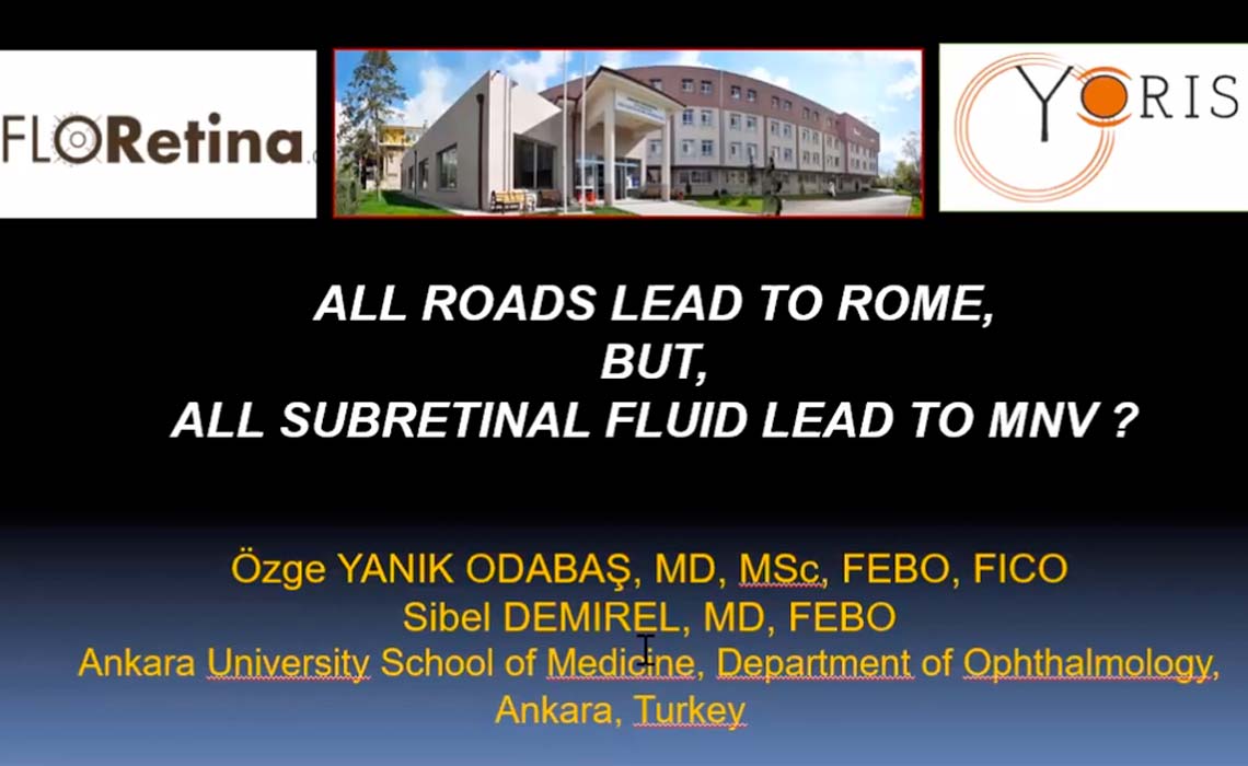
All roads lead to Rome, but, all subretinal fluid lead to MNV?
Ozge Yanik Odabas Sibel DemirelA sixty-eight-year-old female presented with minor visual disturbances in her both eyes. Structural B-scan OCT revealed drusenoid PEDs with overlying hyperreflective material in the right eye, and hyperreflective foci located in the inner retinal layers of left eye. Fundus autofluorescence showed hyperautofluorescent dots and areas in both eyes. During follow-up period, the imaging findings of the macular lesions start to resemble butterfly shaped pattern dystrophy with linear pattern characteristic, and subretinal fluid was developed. However, an MNV lesion did not exist on dye angiography. This case highlights the coexistence of non-neovascular AMD, acquired vitelliform material, and subretinal fluid with extraordinary evolution of these retinal lesions. Subretinal fluid and acquired vitelliform material may develop during the follow-up of non-neovascular AMD cases.
Watch now© 2026 Copyright:
Floretina.com | P.I.
01477560138
Privacy Policy
|
Cookie Policy
|
Terms and Conditions

